
Fasciculus:Autophagy diagram PLoS Biology.jpg
Mensura huius perspectionis: 441 × 599 elementa imaginalia. Aliae mensurae: 177 × 240 elementa imaginalia | 353 × 480 elementa imaginalia | 1 006 × 1 366 elementa imaginalia.
Sua resolutio (1 006 × 1 366 elementa imaginalia, magnitudo fasciculi: 631 chiliocteti, typus MIME: image/jpeg)
Summarium
| Descriptio |
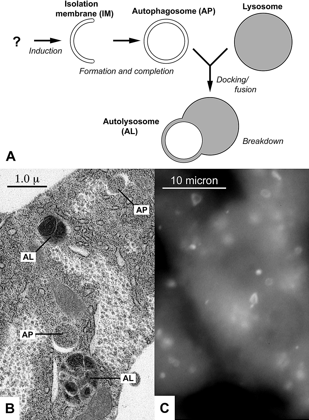
English: Diagram and images of autophagy from a 2006 PLoS Biology review. (A) Diagram of autophagy; (B) Electron micrograph of autophagic structures in the fatbody of a fruit fly larva; (C) Fluorescently labeled autophagosomes in liver cells of starved mice. |
| Datum | |
| Fons | Autophagy: A Forty-Year Search for a Missing Membrane Source. PLoS Biol 4(2): e36. doi:10.1371/journal.pbio.0040036 |
| Auctor | Juhasz G, Neufeld TP. Original images by Ryan Scott (B) and Dr. Noboru Mizushima (C). |
| Permissio (Reusing this file) |
http://www.plosbiology.org/static/license.action |
| Other versions | File:Autophagy diagram PLoS Biology.tif |
Potestas usoris
This file is licensed under the Creative Commons Attribution 2.5 Generic license.
- Tibi licet:
- communicare – copiare, distribuere et committere hoc opus
- to remix – to adapt the work
- His condicionibus:
- attributio – You must give appropriate credit, provide a link to the license, and indicate if changes were made. You may do so in any reasonable manner, but not in any way that suggests the licensor endorses you or your use.
Historia fasciculi
Presso die vel tempore fasciculum videbis, sicut tunc temporis apparuit.
| Dies/Tempus | Minutio | Dimensiones | Usor | Sententia | |
|---|---|---|---|---|---|
| recentissima | 23:55, 3 Septembris 2010 | | 1 006 × 1 366 (631 chiliocteti) | Genericuser | {{Information |Description={{en|1=Diagram and images of autophagy from a 2006 PLoS Biology review. (A) Diagram of autophagy; (B) Electron micrograph of autophagic structures in the fatbody of a fruit fly larva; (C) Fluorescently labeled autophagosomes in |
Nexus ad fasciculum
Ad hunc fasciculum nectit:
Usus fasciculi per inceptus Vicimediorum
Quae incepta Vici fasciculo utuntur:
- Usus in ar.wikipedia.org
- Usus in az.wikipedia.org
- Usus in bn.wikipedia.org
- Usus in bs.wikipedia.org
- Usus in ca.wikipedia.org
- Usus in cs.wikipedia.org
- Usus in de.wikipedia.org
- Usus in en.wikipedia.org
- Usus in en.wiktionary.org
- Usus in es.wikipedia.org
- Usus in fa.wikipedia.org
- Usus in gl.wikipedia.org
- Usus in hy.wikipedia.org
- Usus in id.wikipedia.org
- Usus in kk.wikipedia.org
- Usus in ko.wikipedia.org
- Usus in mk.wikipedia.org
- Usus in nl.wikipedia.org
- Usus in ru.wikipedia.org
- Usus in ru.wikinews.org
- Usus in ru.wiktionary.org
- Usus in simple.wikipedia.org
- Usus in sr.wikipedia.org
- Usus in ta.wikipedia.org
- Usus in te.wikipedia.org
- Usus in zh.wikipedia.org