
Fasciculus:A computed tomography brain scan showing bilateral basal ganglia calcification.jpg

Mensura huius perspectionis: 800 × 589 elementa imaginalia. Aliae mensurae: 320 × 235 elementa imaginalia | 640 × 471 elementa imaginalia | 1 024 × 753 elementa imaginalia | 1 200 × 883 elementa imaginalia.
Sua resolutio (1 200 × 883 elementa imaginalia, magnitudo fasciculi: 153 chiliocteti, typus MIME: image/jpeg)
| Descriptio |
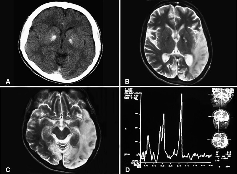
English: (a) A computed tomography brain scan showing bilateral basal ganglia calcification; the cerebellum shows prominent folia indicating mild cerebellar atrophy. (b) Axial T2 brain magnetic resonance image scan showing left temporo-parieto occipital ischemic lesion. (c) Axial T2 brain magnetic resonance image scan showing the extension of the parietal temporal region to the occipital lobe, and also showing a right occipital lesion. (d) Magnetic resonance spectroscopy showing inversion of J-coupling phenomenon at 1.3 ppm, indicating lactate peak. Abu-Amero et al. Journal of Medical Case Reports 2009 3:77 doi:10.1186/1752-1947-3-77 |
| Datum | |
| Fons | A patient with typical clinical features of mitochondrial encephalopathy, lactic acidosis and stroke-like episodes (MELAS) but without an obvious genetic cause: a case report |
| Auctor | Abu-Amero KK, Al-Dhalaan H, Bohlega S, Hellani A, Taylor RW. |
| Permissio (Reusing this file) |
© 2009 Abu-Amero et al; licensee BioMed Central Ltd. This is an Open Access article distributed under the terms of the Creative Commons Attribution License (https://creativecommons.org/licenses/by/2.0), which permits unrestricted use, distribution, and reproduction in any medium, provided the original work is properly cited. |
This file is licensed under the Creative Commons Attribution 2.0 Generic license.
- Tibi licet:
- communicare – copiare, distribuere et committere hoc opus
- to remix – to adapt the work
- His condicionibus:
- attributio – You must give appropriate credit, provide a link to the license, and indicate if changes were made. You may do so in any reasonable manner, but not in any way that suggests the licensor endorses you or your use.
Historia fasciculi
Presso die vel tempore fasciculum videbis, sicut tunc temporis apparuit.
| Dies/Tempus | Minutio | Dimensiones | Usor | Sententia | |
|---|---|---|---|---|---|
| recentissima | 10:20, 28 Ianuarii 2010 | | 1 200 × 883 (153 chiliocteti) | CopperKettle | {{Information |Description={{en|1=(a) A computed tomography brain scan showing bilateral basal ganglia calcification; the cerebellum shows prominent folia indicating mild cerebellar atrophy. (b) Axial T2 brain magnetic resonance image scan showing left te |
Nexus ad fasciculum
Ad hunc fasciculum nectit:
Usus fasciculi per inceptus Vicimediorum
Quae incepta Vici fasciculo utuntur:
- Usus in ar.wikipedia.org
- Usus in de.wikipedia.org
- Usus in en.wikipedia.org
- Usus in en.wikibooks.org
- Usus in es.wikipedia.org
- Usus in fa.wikipedia.org
- Usus in fr.wikipedia.org
- Usus in it.wikipedia.org
- Usus in ru.wikipedia.org
- Usus in sv.wikipedia.org
- Usus in tr.wikipedia.org
- Usus in www.wikidata.org
- Usus in zh.wikipedia.org